We’ve long known that every cancer tumor starts as an abnormality in a single cell but how that one cell progresses to a tumor isn’t well understood. A testing technique that combines specialized microscopy with artificial intelligence provides new insights on how breast cancer tumors develop.
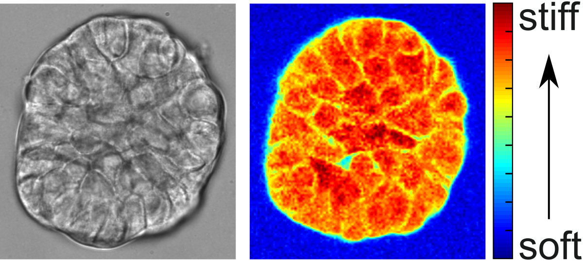
A team led by Jitao Zhang, assistant professor in the Department of Biomedical Engineering, used Brillouin microscopy — a non-invasive mechanical imaging method — to study breast cancer cells in their native 3D context.
“We tracked the stiffness changes of cancer cells for eight days as they grew from single cells into a cluster known as a spheroid,” Zhang said. “This is the first time that the mechanical evolution of cancer spheroids during early growth has been captured.”

In addition to helping cancer biologists better understand the early progression of tumors, this work improves the classification accuracy of breast cancer cells from 74% to 95% after incorporating newly acquired mechanical data.
Brillouin microscopy is an emerging technology that complements existing mechanical testing tools. Zhang’s team advances its power by combining the data from mapping cancer cells with a machine learning algorithm and a deep learning pipeline to streamline the process of data analytics.
Knowledge gained from Brillouin-based studies can inform the development of new diagnostic and therapeutic strategies for diseases such as metastatic cancers, birth defects, and ovarian aging.
To learn more about Zhang’s research, visit:
MSU College of Engineering Media and Public Relations page