
Diagnosing and treating disease is often a long and uncertain process. Symptoms can take years to explain, therapies may not work the same way for every patient, and doctors frequently lack tools that reveal what’s happening inside the body early enough to change outcomes.
At Michigan State University, researchers are working to change that timeline.
Across campus, scientists, physicians, engineers and veterinarians are using advanced imaging systems, radiopharmaceuticals and molecular technologies to develop more precise ways to detect, monitor and treat disease in both animals and humans.
This work is powered by world-class research infrastructure found at only a handful of institutions nationwide. Together these facilities exemplify MSU’s “One Team, One Health” vision — a collaborative and interdisciplinary approach that brings together expertise from various fields that include human medicine, veterinary medicine, agriculture, plant science, environmental science and engineering. This collaboration across disciplines recognizes the interconnectedness across human, animal and environmental health in efforts to solve some of today’s most complex health challenges.
Seeing disease earlier and more clearly
Inside the 13,000-square-foot Center for Imaging and Image-Guided Therapies, or CIIGT, in MSU’s Biomedical Research Complex, researchers have started using a brand-new imaging system that can capture anatomical structures and biological activity simultaneously, faster and in better quality. This leads to a more complete picture of disease, which can aid early detection, diagnosis and monitoring. As a place for scientific discovery, CIIGT helps create new approaches to treating disease, ultimately guiding improved patient care.
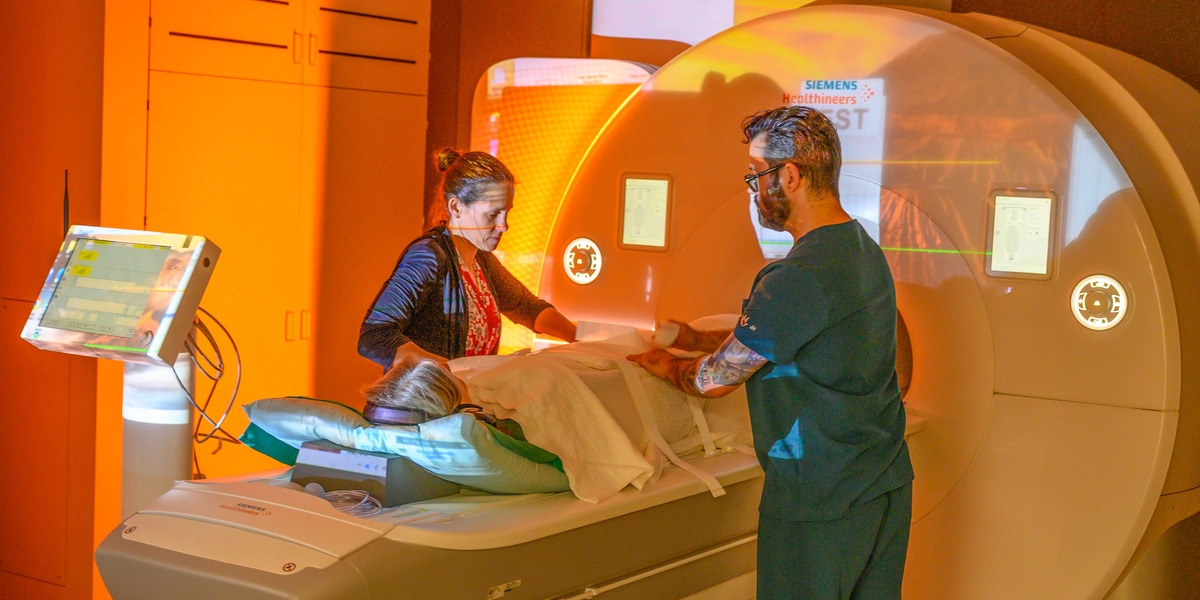
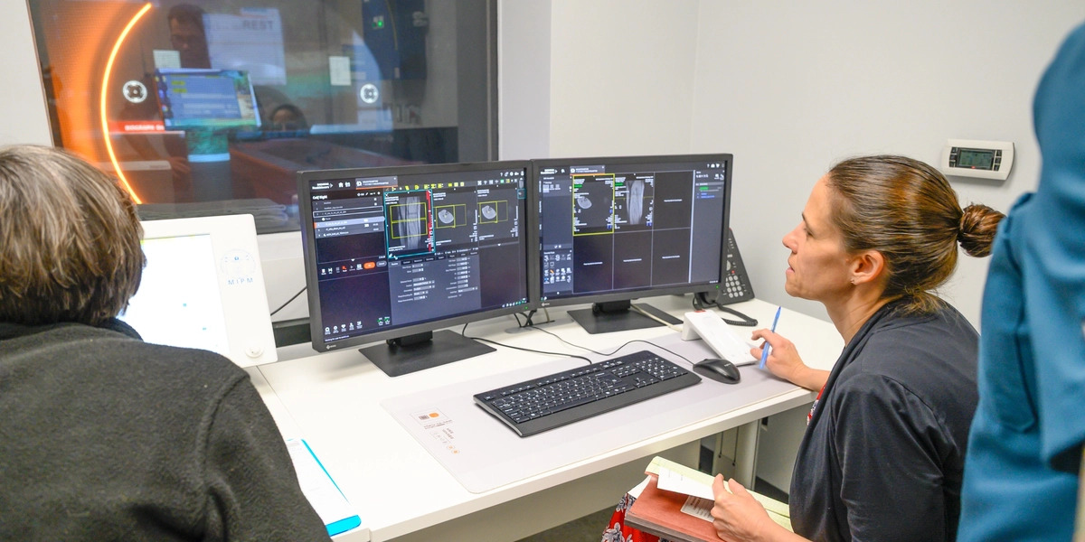
The 9-ton Siemens Biograph One scanner, installed in December 2025, is one of only a handful in the United States. It combines magnetic resonance imaging, or MRI, with positron emission tomography, known as PET, into one simplified, integrated process.
“It not only sees where the disease is, but it can see what’s happening biologically,” says Mark DeLano, professor and chair of the Department of Radiology, based in the College of Human Medicine and the College of Osteopathic Medicine at MSU.
Researchers from across campus will use the device to study brain and cardiovascular function in general, but also diseases ranging from Alzheimer’s and Parkinson’s to cancer.
The applications are wide-ranging. For example, the College of Music has done functional brain imaging to study the perception of music by observing how the brain response of a musician differs from that of a nonmusician when listening to Mozart. More recently, David Kaufman’s research on mild traumatic brain injury in Spartan football players has made use of functional MRI scanning and radiographic biomarkers to assist with prognosis.
While the PET component is currently awaiting approval for human use, researchers are already using the Biograph One’s MRI capability to do advanced PET and MRI simultaneous scanning to study animal models of disease and MRI in human studies, like Jill Slade McMahon’s work investigating oxygen supply to muscles in hypertension and how exercise can help.
Because MSU is home to Michigan’s only veterinary medicine college, the facility offers a unique opportunity to make connections between small animal and larger animal research that could inform the development of new healthcare pathways.
“Its scope covers much ground, driving innovations in medium-to-large animal models, veterinary medicine as well as research focused on Alzheimer’s disease and related dementia, Parkinson’s disease, mild traumatic brain injury and human cancer research with unprecedented imaging capabilities,” says Norman Scheel, an assistant professor in the Department of Radiology.
And that’s exactly how the centerwas designed to operate: as a multidisciplinary facility where both comparative research (which studies animal models alongside human disease) and translational research (which aims to quicken the path of biomedical research findings to a clinical setting) take place.
The renovation and development of CIIGT was made possible by a $6.7 million National Institutes of Health grant, secured by Anna Moore, principal investigator, associate dean for research and development, director of the Precision Health Program and professor in the Departments of Radiology and Physiology. As a crucial part of establishing CIIGT, the cutting-edge Biograph One from Siemens — an MSU partner and industry collaborator — brings MSU’s strengths in collaborative research together, with the ultimate goal of facilitating a smoother pathway from discovery to patient care.
Delivering more targeted treatments
If the Biograph One helps researchers see disease more clearly, RadCore is helping them treat it more precisely.
At RadCore, the Radiochemistry and Radiopharmacy Solutions facility also located in MSU’s Biomedical Research Complex, researchers are focused on developing radiopharmaceuticals that can be used to precisely locate diseases like cancer and neurological conditions and deliver targeted medical treatments.
This approach is part of an emerging medical frontier called theranostics, which fuses diagnostic imaging with targeted therapies using radioactive drugs known as radiopharmaceuticals. Though RadCore occupies a compact 550-square-foot radiopharmacy, the facility is chock full of equipment to manufacture these drugs that are used in nuclear medicine for both diagnostic imaging and treatment of diseases.
“This facility is unique in that it’s very flexible — it does what we need for research purposes, for clinical trials and for therapeutic applications in both veterinary and human medicine,” says Kurt Zinn, professor in the departments of Radiology, Biomedical Engineering, Pharmacology and Toxicology, and Small Animal Clinical Sciences and director of RadCore.
Working with "hot cells", or heavily shielded containment chambers lined with lead and the two cyclotrons behind them, researchers can manufacture radioactive tracers, which facilitate more precise medical imaging that results in better targeted treatments.
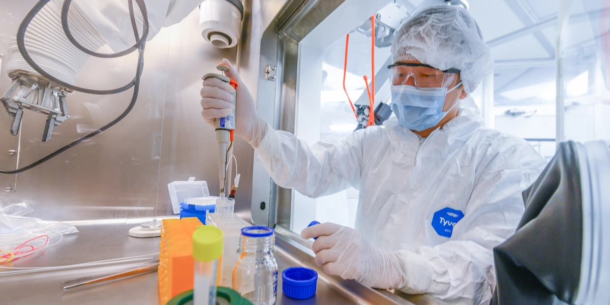
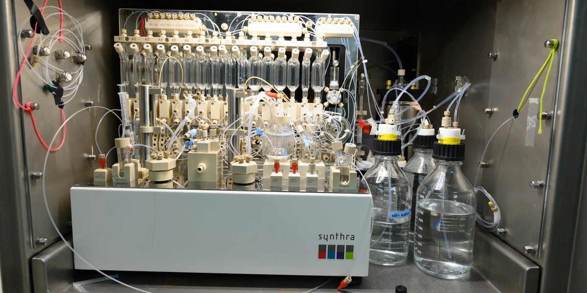
“Different neurological disorders — and even different forms of dementia — can arise from different underlying pathological processes,” says Jinda Fan, an assistant professor in the departments of Pharmacology and Toxicology, Chemistry and Radiology who leads radiochemistry and molecular imaging efforts at RadCore.
Researchers and physicians use radiotracers to visualize these processes by imaging specific molecular markers, which helps them better understand the underlying source of a patient’s symptoms, guiding more targeted treatments and minimizing damage to healthy tissue.
Currently, RadCore produces fluorine-18–labeled radiotracers that are widely used in cancer imaging. Future expansion plans include the production of radiotracers for Alzheimer’s disease and other neurological disorders.
The final drug products are prepared, quality tested and packaged in a current good manufacturing practice, or cGMP, cleanroom in the Radiopharmacy, then immediately available for use with the Biograph One or transported to MSU’s colleges of Human Medicine and Veterinary Medicine. Radiopharmaceuticals are also distributed locally for clinical trials at hospitals in the Lansing area, and production has been expanded to support gene therapy clinical trials as part of the Henry Ford Health + Michigan State University Health Sciences partnership.
Engineering health in nanoscopic detail
Precise treatments depend increasingly on technologies that can deliver them efficiently and are tailored to the needs of each individual patient. At MSU’s Institute for Quantitative Health Science and Engineering, or IQ, researchers are using one of the world’s most accurate 3D bioprinters to create the medical devices and biological structures that can deliver these treatments down to nanoscopic scale.
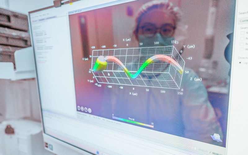
The Nanoscribe Quantum X bio was installed in the IQ 3D Printing Core in 2023 as the first of its kind in the U.S.
As a bioprinter, it can print structures as small as about 150 nanometers — roughly 1,000 times thinner than a human hair — and can even print living cells, opening new possibilities for tissue engineering, disease modeling and personalized medicine. It also prints more rapidly than other microfabrication processes can create, allowing researchers to do iterative work at speed on a range of designs.
“Live-cell printing is challenging because the cells are under stress during the printing process, causing them to lose humidity,” says Yulu Cai, a doctoral candidate in chemical engineering and materials science in the Li lab at the 3D Printing Core. “The Quantum X bio provides a sterile, temperature-controlled environment that’s cell-friendly.”
The technology is already enabling researchers in the Li lab, led by Jinxing Li, Red Cedar Distinguished Assistant Professor in the Department of Biomedical Engineering and faculty director of the IQ 3D Printing Core, to develop next-generation diagnostic and therapeutic devices.
In one project, the Li lab is creating microscopic robotic structures designed to enter tumors and destroy cancer cells with precisely targeted heat, a minimally invasive approach that could one day deliver effective treatment while also reducing damage to surrounding healthy tissue. The work earned the lab an NIH National Institute of Biomedical Imaging and Bioengineering Trailblazer Award in 2025.
Taken individually, each facility represents a major technological investment. Together, they reflect something larger: as a leading public research institution, MSU brings scientists, clinicians and engineers across disciplines together and provides the advanced infrastructure to help bridge the gap between scientific discovery and patient care.
For generations, Spartans have changed lives through research and innovation. Support from federal, state and local funding helps power discoveries that improve health, strengthen communities and keep America at the forefront of innovation and competitiveness. From lifesaving cancer treatments to advances in agriculture, energy and technology, see how Michigan State University researchers are shaping a better future for Michigan and the world.
Story courtesy of MSUToday.
MSU College of Engineering Media and Public Relations page